


Disclaimer: This article is intended for educational and research purposes only. It is NOT medical advice. Do not attempt anything you read without medical supervision.
If you wanted everyone tired, inflamed, and begging for a prescription, there’s one organ you’d want Memory Holed.
The thymus trains T cells to spot infection and developing cancers without destroying your own tissue. It coordinates repair and manages inflammation too.
It starts shrinking when you’re a teenager. By 40, it’s almost gone.
Your doctor was taught this is fine (same curriculum teaching DHT is useless after puberty).
Colds become “the man flu”, a workout is 4 days of crippling DOMs, and brain fog becomes default mode, just slow enough for you to not notice.
The doctor calls this “aging”. Convenient word when no one can identify a cause or solution.
Then someone sells you elderberry gummies because the whole world still thinks immune function is simply “fighting germs”. Lmao.
The deeper problem is rebuilding the thymus between insults. That is the Thymic Repair Bottleneck.
That’s where a Russian Peptide used by Olympic Athletes, Elite Military, and Cosmonauts, looks promising. The peptide of interest? Vilon.
The following guide was written with Love on 2mg :)
What Vilon is
The molecule contains two amino acids. K is lysine. E is glutamic acid. They are joined in that order.
The Soviet naming department still found a way to make this confusing.
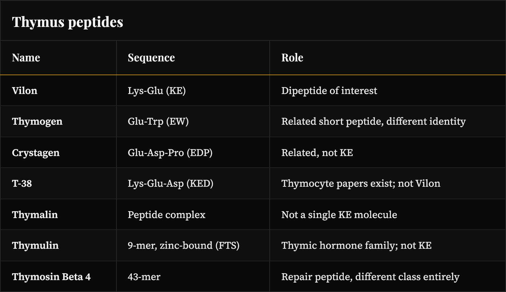
The bottle says Vilon. Its government name is KE.
That gives KE the molecular formula C₁₁H₂₁N₃O₅ and a molecular weight around 275 g/mol.
Simple enough, until reading the thymic peptide feels like a CAPTCHA:
EW is Thymogen, glutamic acid followed by tryptophan.
EDP is Crystagen, Glutamic acid–Aspartic acid–Proline
KED (Vilon + Aspartic Acid) is Vesugen, the vascular and endothelial bioregulator.
Thymalin is the whole thymic peptide complex. It contains Vilon plus other thymic peptides.
Thymulin is a separate 9-amino-acid peptide.
KE contains only two amino acids, yet it accumulated decades of receipts.
Most sit inside Khavinson’s thymic bioregulator literature, where short peptides were studied as signals for aging organs losing capacity.
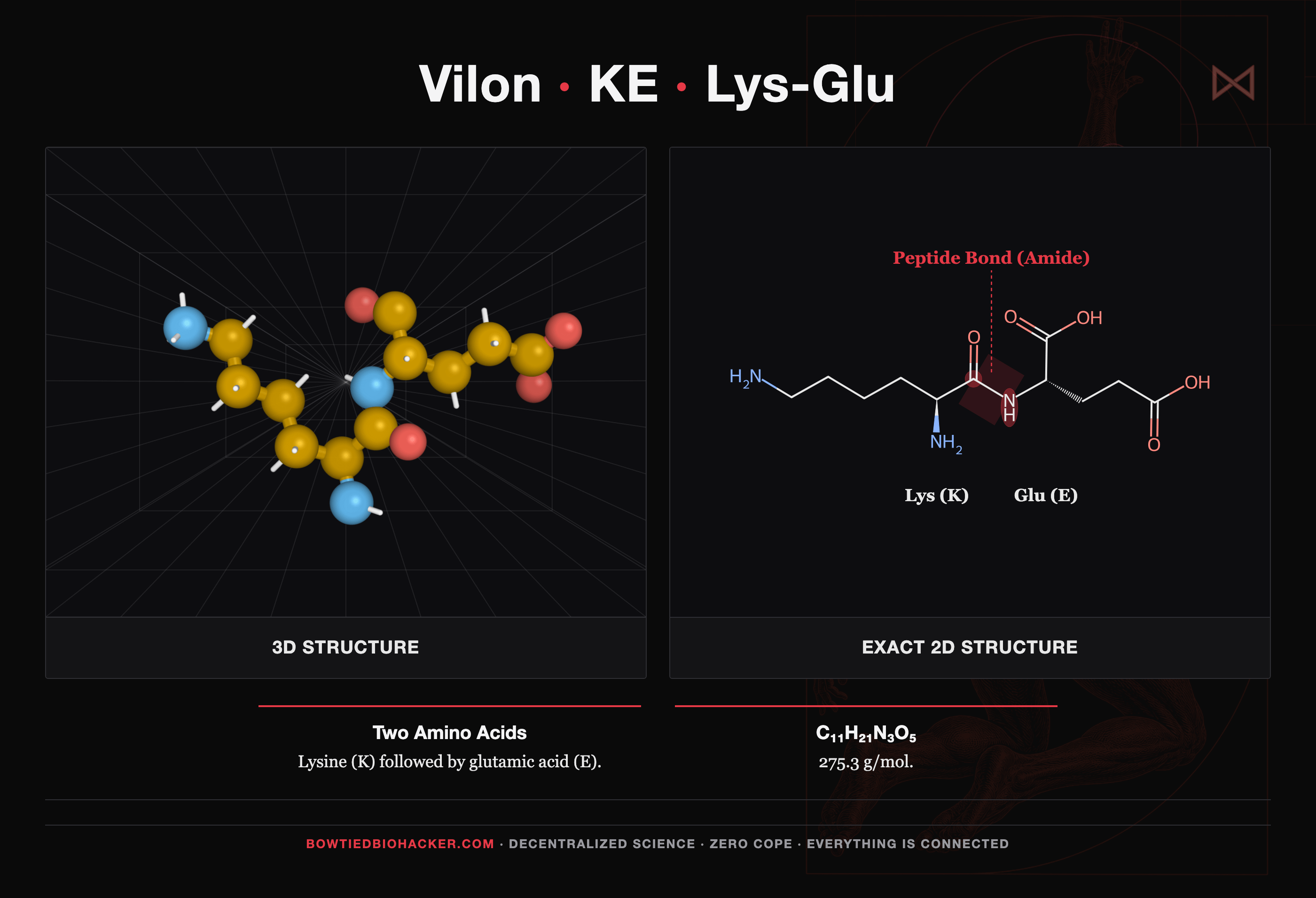
Lysine joined with glutamic acid. Two amino acids and nothing else.
The bad week becomes your baseline
Everyone reading this has a number they avoid calculating.
How many peak years do you have left, and which birthday comes with the next real “downgrade”?
How much repair capacity you retain shapes that timeline. Repair capacity is also the one thing nobody sells you.
A session feels great while you are training, then leaves you wrecked for days. The same bounceback used to take less than 24 hours.
The COVID era left a lot of people with disrupted T cells and persistent inflammation. Most feel like they never got their full edge back.
High inflammatory tone takes away the mostv and gives you the least to point at. Your face looks puffy in the morning. Your joints need an hour to loosen. The afternoon goes flat regardless of sleep, and your skin looks “tired” in pictures.
You can explain away every one of those things.
That works until you realize you have been explaining away the same bad week for years.
Open the Vilon papers and the same biology keeps appearing: thymocytes, thymic epithelium, chromatin opening in elderly lymphocytes, interleukin 2 gene expression, macrophage cytokine tone, and coagulation and fibrinolysis markers.
Every item on that list is a repair dial.
This is the system that rebuilds tissue, resets inflammatory tone, and decides how much capacity you have left after 30.
In a 2021 interview, Khavinson discussed how Soviet Olympic athletes began using his peptide bioregulators in 1980. Soccer players, tennis players, and track athletes followed, mainly for recovery before and after competition. He also mentioned cosmonauts, submariners, and elite military units used similar protocols. (Aging Matters interview with Khavinson, 2021) Seems like extended rest days were not acceptable in Soviet performance plans. Good thing that’s where Vilon stands out from other peptides.
What People Report On Cycle
Research groups tell us what moved. Training logs and careful n=1 runs tell us what it felt like.
The reports tend to arrive in the same order.
Soreness usually moves first. Sessions that caused several days of soreness clear faster, and people tracking a cycle report noticing this during the first days.
The effects usually deepen as the cycle continues. Training density and autonomic markers keep improving as the weeks pass.
Work capacity holds longer before quality falls off. People report more endurance and more training volume without feeling wrecked afterward.
People also describe clean energy and a general sense of well being: more alertness and awareness, less brain fog, and the feeling that the sludge has cleared out of the body. Physical work can feel less painful or even good.
Resting heart rate tends to fall rather than climb, so the energy does not feel driven by a stimulant. Some users report higher HRV as recovery improves.
Sleep and recovery improve together. Deeper nights usually travel with the same HR shift.
Some people report insomnia when they take Vilon late, so keep it to the morning.
Where Vilon comes from
Thymosin β4 (TB4, AKA what most people’s “TB-500” actually is) is a 43 amino acid peptide found in the thymus, platelets, and injured tissue. The human TB4 sequence contains a KE pair within the PSKET region.
TB4 carries other short bioactive stretches people already know by other names, including the SDKP motif and the LKKTETQ actin binding region that commercial TB500 fragments are built around. KE is liberated when longer thymic peptides get broken down by proteases in tissue, blood, or gut.
Eating thymus is microdosing Vilon
Whole thymus tissue has more peptides than your local Bodega: TB4, Ta1, Thymulin, the short fragments those proteins break off into, including free and bound Vilon. Calf sweetbreads, thymus meat, and the desiccated thymus extracts on supplement shelves all deliver the same thing in uneven and unlabeled amounts.
This is microdosing Vilon the ancestral way, though don’t expect the same effects of 1mg+ Vilon in enteric capsules.
Note: this may actually be the way to go as something you use daily or to maintain results from a Vilon cycle.
The COVID Question Worth Asking
COVID took plenty of healthy people and left them as chronically fatigued shells of their former selves.
The mRNA jabbed were not spared either (that group usually brings more issues).
Across the larger COVID studies, the same two problems keep appearing:
Persistent T cell dysfunction.
Inflammatory and clotting pathways that refuse to shut off.
Vilon’s published work covers thymus and T cell biology, coagulation, and fibrinolysis. That overlap gives researchers a clear reason to investigate Vilon as an adjunct therapy.
Then Thymalin makes the question much harder to ignore.
Mass spectrometry and HPLC showed that Thymalin contains Vilon, Thymogen, Crystagen, and other peptides (Khavinson et al., Biological Bulletin Reviews, 2021).
Thymalin was then tested in a single-blind randomized controlled trial involving older patients with severe COVID. These patients were on oxygen with roughly half their lung tissue affected.
Hospital mortality fell from 40.9% to 19.4%.
Better than half.
The immune markers were even crazier:
Blood lymphocytes increased 92%.
T cells increased 2.2 times.
NK cells increased 2.4 times.
CD4+ and CD8+ cells each increased 2.2 times.
Interleukin 6 fell 6.5 times.
C reactive protein fell 3.3 times.
D dimer decreased.
Standard therapy moved none of those markers (Kuznik, Khavinson et al., Advances in Gerontology, 2021).
The overlap is still a NO BRAINER research question.
Who This Is For
Look into Vilon if:
You train hard and care about recovery quality.
You want more weekly volume without extended soreness and a high resting heart rate.
You already use TRT or exogenous androgens and want a repair layer that may add some protection. Testosterone, estradiol, and cortisol can accelerate thymic involution (Sutherland et al., Journal of Immunology, 2005; Madan et al., Journal for ImmunoTherapy of Cancer, 2013).
You cannot afford to spend the workweek recovering from the gym.
You are detrained or dealing with exercise intolerance, chronic fatigue, or hypoxia.
You already handle sunlight, sleep, and nutrition and want to investigate the thymus side of aging.
You are tired of being a pin cushion but still love HIGH ROI performance-enhancing compounds.
You are losing hair.*
*We have not compared notes with anyone balding at a concerning rate. HOWEVER, the hypoxia and immune modulating mechanisms make this worth investigating. Some people have reported “lots of baby hairs growing.” New readers should know that hair loss has been reliably solved in the latest protocol.
Who Should Stay Away
If a qualified professional has examined your situation and told you to stay off Vilon, take that seriously.
Skip it if you are already running a chemistry set of peptides and bioregulators. Best case scenario, you will have no idea what caused what.
The Protocol We Run
Dose: A common oral protocol uses 1 to 4 mg of enteric coated KE per day. 1 mg is a good starting point. Take notes on recovery and response. Our go to source is YourProtocol.co, and BOWTIED gets you 10% off.
Timing: morning or earlier in the day, empty stomach, per product and practitioner guidance.
Cycle: Khavinson research follows short (30 days or less) structured cycles with breaks lasting several months. Is it a good idea to run longer than that? We don’t know.
Track every cycle: resting heart rate, HRV, weekly training volume, soreness, sleep, session quality, and infections.
Get labs. A lymphocyte subset panel with naive markers is the only thing on this list that looks at the mechanism instead of the downstream feel. Draw one before your first capsule and one after the cycle, because a cycle you cannot measure is a cycle you cannot judge.
Recovery may move within days, while resting heart rate, HRV, and volume tolerance can keep improving through the cycle. Judge the trend across the cycle. One freakishly good Tuesday proves nothing.
When readers ask where to get a labeled enteric KE capsule instead of a gray market research vial, this is the one we use: vilON, 1 mg KE, 60 HPMC enteric capsules, rice flour, USA cGMP, independently tested, best in class customer service, and zero reports of substandard quality.
Why Oral Works Best
Most of the time injection is necessary, as oral peptides get destroyed in the gut. While that is true for most larger peptides, KE contains only two amino acids and fits right into the ideal transport proteins.
The intestine has transporters for dipeptides (like Vilon) and tripeptides: PEPT1, encoded by SLC15A1, transports small peptides from the intestinal lumen into enterocytes. PEPT2, encoded by SLC15A2, lives in the kidneys to recycle filtered peptides.
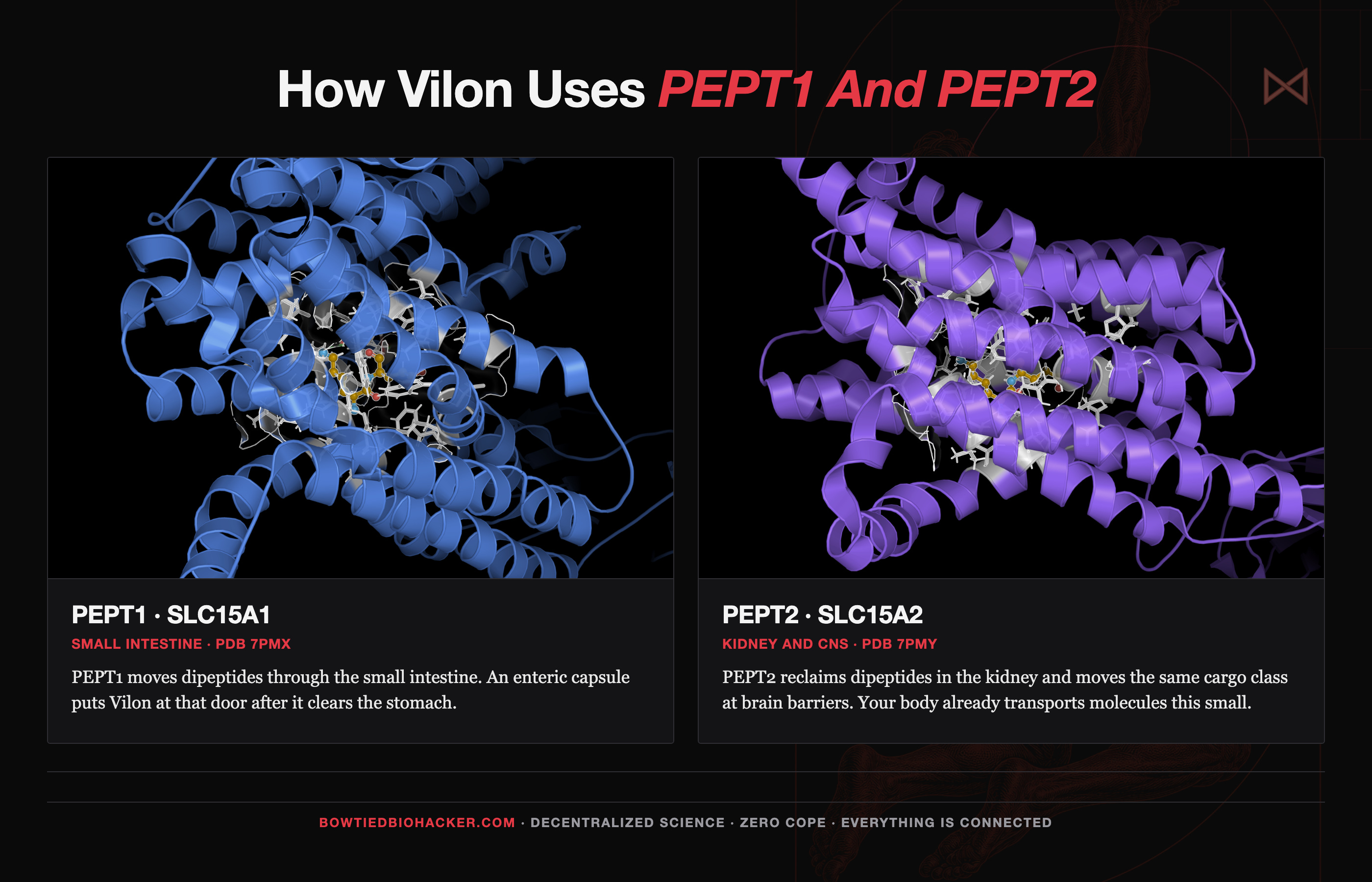
PEPT1 and PEPT2 transport dipeptides and tripeptides across cell membranes. PEPT1 handles intestinal absorption, while PEPT2 helps reclaim filtered peptides in the kidneys. Vilon sits in the pocket where the experimental alanine and phenylalanine dipeptide was crystallized. No Vilon cocrystal exists, so read this as a size match at the published substrate site.
Your gut did not evolve PEPT1 for Russian gerontologists. It exists because every steak, egg, and glass of milk becomes a flood of small peptides during digestion. Mammalian PEPT1 transports molecules like KE across the gut wall (Meredith, 2009), and oral Vilon altered digestive enzyme activity in rats (Khavinson, 2001).
What other published research shows
Blood flow and clotting
In type 1 diabetics with DIC-like clotting dysfunction, Vilon restored antithrombin III and Protein C and moved fibrinolysis toward normal (Kuznik et al., 2006). Nobody measured gym recovery, but better clot regulation could support microvascular oxygen delivery and metabolite clearance, giving the athlete reports a plausible mechanism.
Repair and regeneration start in the thymus
The strongest thymus evidence hits the right cell type directly. In a 2004 study, Vilon increased nucleolar organizer region activity in thymocytes and thymic epithelial cells. It also increased blast transformation in thymocytes, indicating that more of the cells had entered an activated, proliferative state (Raikhlin 2004). The epithelial cell finding matters because thymic regeneration depends on restoring the stromal environment that supports T cell development. The study didn’t assay FOXN1, so the honest reading is that Vilon activates the right cell class.
The rest of the published work on KE fills in the picture.
In rats, Vilon stimulated thymocyte proliferation, thymocytes being T cells still in training, and helped radiosensitive organs recover after radiation exposure (Khavinson et al., Bull Exp Biol Med 2001).
It amplified the comitogenic signal that pushes thymocytes to divide, modulated interleukin 1 beta comitogenicity, and shifted sphingomyelinase pathway activity in thymocyte membranes (Khavinson et al., 2002).
Vilon shifted CD4 and CD5 expression in thymic cells toward a T helper phenotype (Sevostyanova et al., 2013; reviewed in PMC8365293).
In spleen cells, it induced interleukin 2 mRNA without the usual external stimulation, suggesting a direct effect on gene regulation rather than a nonspecific stress response (Kazakova et al., 2002).
Russian gerontology studies also report partial protection against radiation associated aging of the thymus and spleen, along with reduced thymic involution under stress.
Add those up and the anti aging case is the deepest thing on offer here: rebuild the thymic epithelial compartment, push T cell precursors to mature, and keep that tissue working under stress.
How Vilon modulates inflammatory signaling
The most useful modern paper is Avolio et al., IJMS 2022. The researchers tested Vilon, labeled P2 in the paper, in human monocyte and macrophage cell lines. The study measured how the peptide changed inflammatory signaling inside human immune cells. Four results stand out:
In differentiated macrophages, Vilon increased STAT1 phosphorylation through what appeared to be a receptor independent mechanism.
Its effects on ERK and JNK depended on the state of the cell, which suggests immune MODULATION rather than stimulation or suppression.
The peptide panel containing Vilon also reduced TNF, interleukin 6, and interleukin 17 after LPS exposure. LPS is bacterial endotoxin commonly used to provoke an inflammatory response in cell experiments.
Most peptides tested turned down adhesion molecules (the surface tags immune cells use to stick to blood vessel walls and crawl into tissue). Less sticking = less inflammatory accumulation.
Nobody tracked soreness or HRV because the subjects were macrophages in a dish. The study still gives us something valuable: a direct measurement of inflammatory signaling.
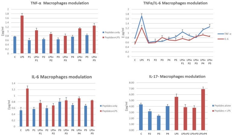
From Avolio et al., IJMS 2022: short Khavinson peptides, Vilon among them, run through inflammatory readouts after stimulation with LPS in macrophages.
Chromatin and gene expression
In lymphocytes from older donors, KE shifts chromatin toward the working state, with more actively transcribed euchromatin and less condensed heterochromatin (Biol Bull Rev 2021).
KE binds selectively to double stranded DNA, with reported affinity for the TCGA sequence (PMC8365293; broader peptide and gene review PMC8619776).
In aging human mesenchymal stem cells, KE altered the expression and protein levels of SIRT1, PARP1, and PARP2, all of which participate in DNA repair and cellular aging. Molecular modeling identified GCGG and GGGC promoter sequences as possible binding sites (PMID 37782636).
Reviews suggest effects on IGF1, FOXO1, TERT, TNKS2, NFκB, cytokine regulation, and fibrinolysis in aging cell models.
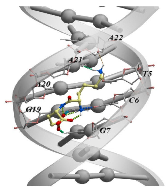
From a 2021 review of short peptide DNA interactions: how a molecule this small can sit down on DNA and chromatin and change which genes get read. The figure shows the mechanism class for Khavinson peptides as a group.
The vascular, thymic, and inflammatory findings come from different experiments. Gene regulation is one possible link between them, especially because KE has reported interactions with DNA and several aging related genes. The thymus connection brings one transcription factor into focus.
The gene and promoter map
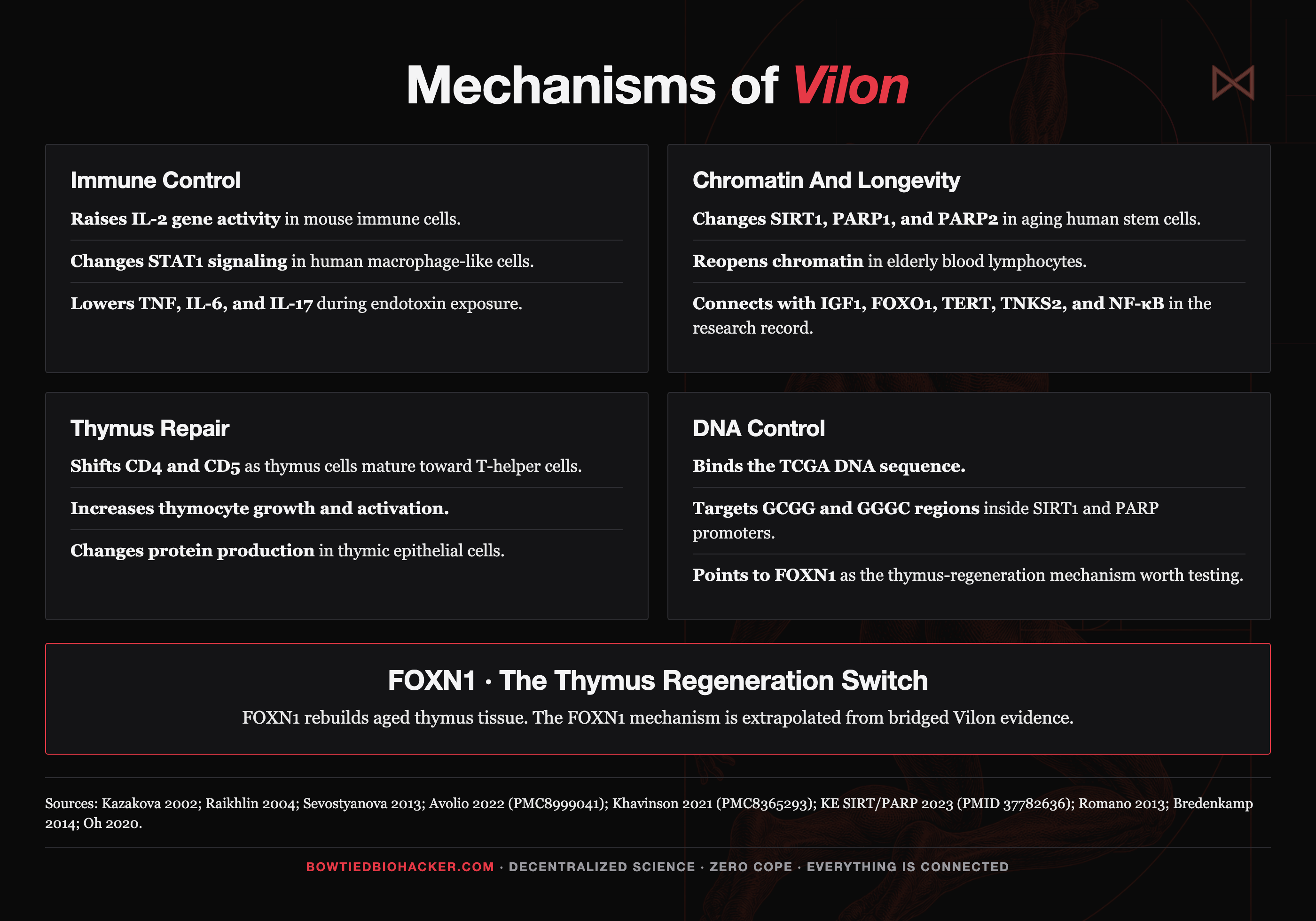
Genes and promoter motifs associated with KE. Solid nodes carry direct or nearly direct expression evidence. Softer nodes are extrapolations from review papers.
FOXN1, the regeneration gene behind the anti aging case
Real thymic rejuvenation runs through FOXN1, the transcription factor that keeps thymic epithelial cells doing their job. Vilon acts on that epithelial compartment, and the changes it produces are consistent with thymus regeneration.
Rejuvenating a thymus is a thymic epithelial cell matter
Thymic epithelial cells create the environment where T cell precursors enter, develop, and undergo selection. Aging damages this stromal scaffold, which explains why supplying young bone marrow cells doesn’t fully restore an old thymus. New progenitors still need a functional thymic environment to mature (Romano 2013; Liang 2022).
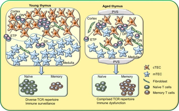
From Liang et al., Aging Cell 2022. As the years climb, the thymus stroma breaks down and the output of fresh T cells falls with it.
Why FOXN1 is needed
Take FOXN1 away and the thymus never finishes building itself. FOXN1 controls several parts of thymic epithelial cell function (Romano 2013; Vaidya 2016):
CCL25 and CXCL12 recruit T cell precursors into the thymus.
The Notch ligand that instructs those arrivals to become T cells (DLL4).
The growth factor support that keeps the nursery running (SCF).
The selection machinery cortical epithelial cells use to test each new T cell before release (β5t/PSMB11 and the MHC class II module).
FOXN1 controls all of it, which is why the thymus cannot be rebuilt without restoring it first.
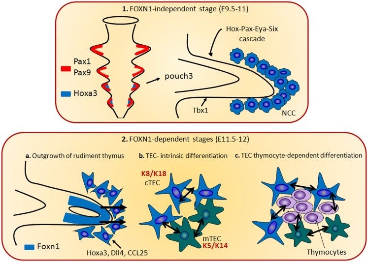
From Romano et al., Front Immunol 2013. The construction blueprint of the thymus, phase by phase. Every phase shown stalls without FOXN1.
Turning FOXN1 back on rebuilds an aged thymus
Increasing FOXN1 specifically in thymic epithelial cells regenerates the aged thymus and starts putting out naive T cells again (Bredenkamp 2014).
Embryonic fibroblasts reprogrammed with FOXN1 and transplanted into old mice produce thymic regrowth, with lower inflammaging signals alongside it (Oh 2020, JCI Insight).
Mice engineered to overexpress Foxn1 resist involution as they age, and they carry less of the peripheral CD4 memory buildup that marks an old immune system (Zook et al.).
The loss of function experiment closes the loop, because removing Foxn1 degrades the peripheral T cell pool into the pattern of age driven immunosenescence (Foxn1 dosage models).
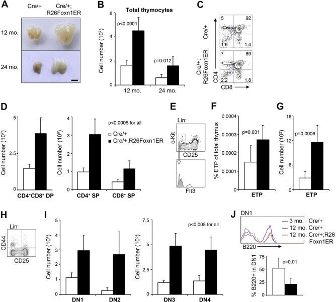
From Bredenkamp et al., Development 2014. The thymus on the left is old and shrunken, the way yours looks after your twenties. The one on the right is the same aged animal after researchers switched FOXN1 back on. The organ regrew.
No study has measured FOXN1 under Vilon. The mechanism remains a strong hypothesis: FOXN1 controls the epithelial compartment Vilon acts on, restoring FOXN1 reverses involution, and Vilon also affects chromatin. The overlap is compelling, but the direct experiment remains undone.
The lab test worth ordering
A standard CBC will not show you any of this. Do not read the lymphocyte to monocyte ratio as a thymus signal, because that ratio moves with inflammation on its own: when monocyte cytokine and STAT1 signaling settles, the number shifts while nothing changes anywhere near the thymus. It works as a rough read on systemic inflammation tone and tells you nothing about whether your T cell pipeline is running younger or older.
For the bloodwork we point people at Vanguard Perform’s Lymphocyte Subset Panel 1. Code BOWTIED for 10% off.
The core panel measures total T cells (CD3), helper and killer T cells (CD4 and CD8), the CD4/CD8 ratio, B cells (CD19), and natural killer cells (CD16/56).
The additional tests below estimate whether the thymus is still shipping fresh naive T cells (as of now, you might need a doctor or clinician to order them, but a private online order should be available soon).
Naive CD4+ and naive CD8+ T cells, gated on CD45RA+CCR7+ or CD45RA+CD62L+ (the fresh cells the thymus has shipped).
sjTRECs on the same draw, if the lab will do it. This is the closest direct readout of thymic output you can get.
Absolute naive counts alongside percentages.
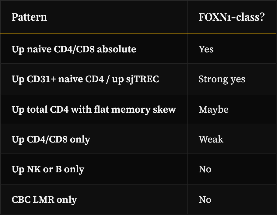
Each subset lined up against the pattern real thymic output would produce. Naive CD4+ and CD8+ up, CD31+ recent thymic emigrant cells up, and sjTRECs up, all in absolute counts, is the combination that points in the direction expected from FOXN1. One marker moving alone does not.
A shift concentrated in naive T cells, with naive rising relative to aged memory cells, is the exact direction thymic restoration predicts. A panel limited to total CD3 cannot see it, so order the fuller subset panel before you draw conclusions.
The experiments worth running ASAP
For a serious n=1, test the subset panel, naive T cells, CD31, and sjTRECs at baseline, weeks 2 to 4, and weeks 8 to 12. Use the same recovery log described above.
The lab experiment that would settle the FOXN1 mechanism is relatively cheap and standard. Drop a comment if you’d donate to help fund it.
Take thymic epithelial cells, expose them to Vilon across a range of doses and times, and measure the chain from gene to protein to target:
FOXN1 messenger RNA (qPCR): Does transcription go up?
FOXN1 protein (immunofluorescence or Western blot): Does it increase?
Downstream targets: CCL25, CXCL12, DLL4, KITL, PSMB11/β5t, and MHC class II should move together.
TEC identity markers: Do the cells remain thymic epithelial cells?
Pass: FOXN1 goes up and at least two of its target genes go up with it.
Fail: FOXN1 stays flat an d the target genes stay flat.
The experiment that would close the gap: knock FOXN1 down in thymic epithelial cells, then add Vilon. If the Vilon response on those same targets dies when FOXN1 is gone, that settles it, because nothing except FOXN1 is left to explain the effect.
Animal experiment: Give Vilon to aged mice and measure thymus cellularity, flow cytometry on the epithelial compartments, a FOXN1 protein stain, and fresh naive T cells in the blood.
References
Romano et al. FOXN1 master regulator. Front Immunol 2013. Full text
Vaidya et al. FOXN1 in thymus organogenesis. Eur J Immunol 2016. Full text
Bredenkamp et al. Regeneration of aged thymus by FOXN1. Development 2014. Full text
Oh et al. fibroblasts reprogrammed with FOXN1. JCI Insight 2020. Full text
Liang et al. Thymic involution with age. Aging Cell 2022. Full text
Zook et al. Foxn1 overexpression and thymic involution. Full text
Foxn1 dosage models, peripheral T cell aging. Full text
Khavinson et al. Vilon/Epithalon radiosensitive organs. Bull Exp Biol Med 2001. Springer
Khavinson et al. Peroral Vilon and digestive enzyme activity. 2001. Springer
Raikhlin et al. NOR proteins in thymocytes and thymic epitheliocytes with Vilon. 2004. PubMed
Khavinson et al. Thymocyte blast transformation / sphingomyelin pathway. 2002. PubMed
Kazakova et al. interleukin 2 gene expression and short peptides. 2002. PubMed
Sevostyanova et al. Immunomodulating effects of Vilon. 2013. PubMed
Khavinson et al. Thymalin / KE molecular aspects. Biol Bull Rev 2021. Full text
Khavinson et al. Peptide regulation of gene expression. Molecules 2021. Full text
Khavinson et al. KE, SIRT1/PARP1/PARP2 in aging MSCs. 2023. PubMed
Avolio et al. Vilon and related peptides in human monocytes and macrophages. IJMS 2022. Full text
Kuznik et al. Vilon, coagulation, fibrinolysis in T1D. 2006. PubMed
Meredith D. Mammalian PEPT1. 2009. Full text
Khavinson interview, peptide bioregulators in sports and space. Aging Matters 2021. Interview
Thymosin β4 sequence context (human Tβ4 / TMSB4X product literature and reviews): KE appears as a contiguous dipeptide in the parent sequence, and SDKP and LKKTETQ are separate known short motifs from the same parent. Sequence presence is chemistry rather than a published isolation paper for commercial Vilon.
Kuznik, Khavinson et al. Peptide drug Thymalin regulates immune status in older patients with severe COVID. Adv Gerontol 2021;11(4):pages 368 to 376. Randomized controlled trial with single blinding, n=36 Thymalin + n=44 control. DOI
Sutherland et al. Activation of thymic regeneration in mice and humans following androgen blockade. J Immunol 2005;175(4):pages 2741 to 2753. PubMed
Madan et al. Impact of androgen deprivation therapy on the thymus and the production of naive T cells. J Immunother Cancer 2013;1(Suppl 1):P83. Full text
Affiliate disclosure: This article contains product links and coupon codes. Some links are affiliate / referral links (including Protocol vilON). We may receive a commission on qualifying purchases made through those links or with codes such as BOWTIED, at no extra cost to you. Proceeds support more research and writing. We only point to products we would run ourselves.
On Monday im doing BW so im adding LYMPHOCYTE SUBET PANEL 1.
Bumped my HRV instantly. Might have flared an auto immune issue but could have just been chance. Really wished I could have taken for longer.